

Another Article About the Neck . . . or Is It?
While the neck is a bridge, a pathway, the position of the neck and head can also indicate a multitude of other things happening beneath the surface.
The infraspinatus is a small, deep muscle located on the posterior surface of the scapula. As its name indicates, it is situated below the spine of the scapula and covers the entire infraspinous fossa. Triangular in shape, the fibers of the infraspinatus are broadest at the medial border of the scapula and converge laterally where they cross the glenohumeral joint, wrap around the humeral head, and attach anteriorly onto the greater tubercle of the humerus. The infraspinatus fascia covers the infraspinatus muscle and separates it from the neighboring teres major and teres minor muscles. The majority of the infraspinatus is framed by the posterior edge of the deltoid, the lateral edge of the lower trapezius, and the superior edge of the latissimus dorsi. The lateral portion is less accessible, as it is obscured by the bulk of the deltoid muscle.
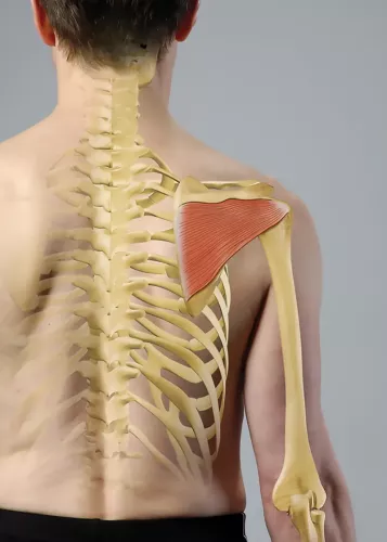
The infraspinatus is one of four muscles that make up the rotator cuff. The supraspinatus, infraspinatus, teres minor, and subscapularis all function as a unit to stabilize the humeral head in the glenoid fossa. Each muscle has a specific role in steering the head of the humerus as the arm moves into different positions. Specifically, the infraspinatus works with the teres minor to seat the humeral head posteriorly in the glenoid fossa and prevent impingement on the coracoid process of the scapula.
The infraspinatus is one of the most powerful external rotators of the glenohumeral joint and is essential in pulling the upper extremity into backward extension and external rotation for shoulder movements such as pitching and hitting overhead. The horizontal and slight upward angle of the muscle fibers, as well as the strong attachment to the posterior scapula, provide excellent mechanical advantage for this "winding up" motion of the shoulder. The infraspinatus is also recruited eccentrically to slow the upper extremity during the "follow-through" or deceleration phase of these powerful movements.
Imbalances often develop between the powerful internal rotators of the shoulder (pectoralis major, latissimus dorsi, teres major, anterior deltoid, and subscapularis) and the smaller external rotators (posterior deltoid, infraspinatus, and teres minor), creating faulty mechanics in the glenohumeral joint. This presents as a variety of pathologies, including impingement syndrome, tendonitis, and bursitis. Trigger points are more common in the deeper subscapularis but may be responsible for referred pain in the upper extremity.
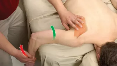
Positioning: client prone with their arm off the edge of the table
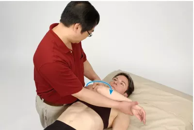
Positioning: client supine at the edge of the table with their arm relaxed at their side
While the neck is a bridge, a pathway, the position of the neck and head can also indicate a multitude of other things happening beneath the surface.
Understanding fibroblasts and the extracellular matrix changes how we think about the tissue we touch.
Studies reveal that 37 percent of the force generated by muscle contraction is transmitted to adjacent connective tissue structures instead of the bones.
Ongoing research suggests the sciatic nerve's healthy functioning depends on its fascial connections.